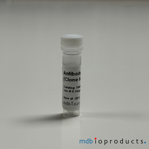
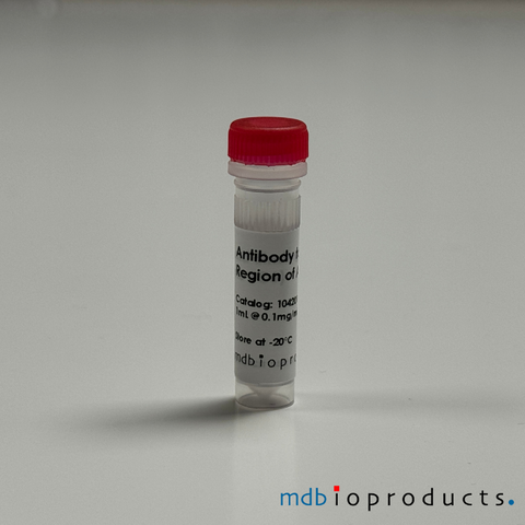
Keratocan Antibody, 100 ug
Keratocan (KERA) monoclonal antibody (mouse, clone Ker-1) to detect keratocan. 1 mL/vial. Concentration is 0.1 mg/mL. Normal keratocan expression in adult tissues is limited to the corneal stroma and is considered...
Antibodies
1042008
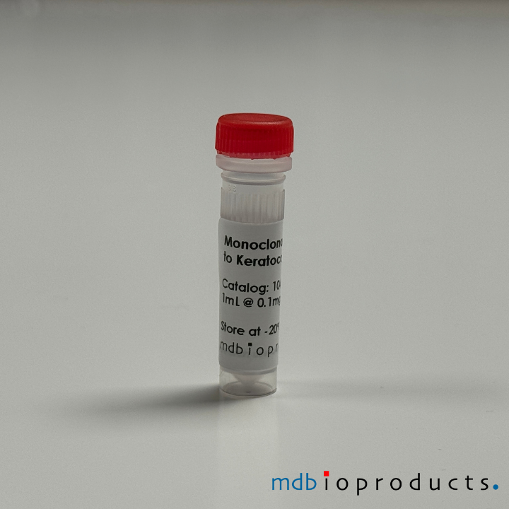
Keratocan (KERA) monoclonal antibody (mouse, clone Ker-1) to detect keratocan. 1 mL/vial. Concentration is 0.1 mg/mL.
Normal keratocan expression in adult tissues is limited to the corneal stroma and is considered a phenotypic marker for keratocytes. In keratocan knockout mice (Kera-/-) the corneal stroma is thinner, the cornea-iris angles are narrower and the collagen fibers of the corneal stroma are disorganized when compared to wild-type animals. In humans, mutations of the keratocan gene (KERA) are associated with the human disease called Cornea plana (CNA2). This disease is characterized by a flattening of the forward convex curvature of the cornea and has been associated with glaucoma. Ultimately, this leads to a decrease in light refraction. Present research is focused on using adult stem cells to regenerate tissue and corneal transparency within knockout mice. Additionally, understanding keratocan in the corneal inflammatory response is a topic of interest.
Related Terms/Symbols:
- KERA
- CNA2
- Keratan sulfate proteoglycans (KSPGs)
- O60938
- KTN
- SLRR2B
References/Citations:
Immunolocalisation and expression of keratocan in tendon. Rees SG, Waggett AD, Kerr BC, Probert J, Gealy EC, Dent CM, Caterson B, Hughes CE. (2008) Osteoarthritis Cartilage.
Fragmentation of decorin, biglycan, lumican and keratocan is elevated in degener- ate human meniscus, knee and hip articular cartilages compared with age-matched macroscopically normal and control tissues. Melrose J, Fuller ES, Roughley PJ, Smith MM, Kerr B, Hughes CE, Caterson B, Little CB. (2008) Arthritis Res Ther. 10(4):R79.
Differential expression of the keratan sulphate proteoglycan, keratocan, during chick corneal embryogenesis. Gealy EC, Kerr BC, Young RD, Tudor D, Hayes AJ, Hughes CE, Caterson B, Quantock AJ, Ralphs JR. (2007) Histochem Cell Biol. Dec;128(6):551-5.
Product Insert (PDF) - Informational use only. Please refer to insert included with product.
Data/Specifications:
- Clone: Ker-1
- Host: Mouse
- Isotype: IgM
- Purity: Affinity purified on thiophilic column
- Form: Liquid, 1 mL/vial
- Concentration: 0.1 mg/mL
- Storage: Store at -20°C.
- Specificity: This Keratocan antibody recognizes a protein core epitope in human, bovine, porcine, mouse & chicken tissues.
Related Products
Additional products you might interested in.
-
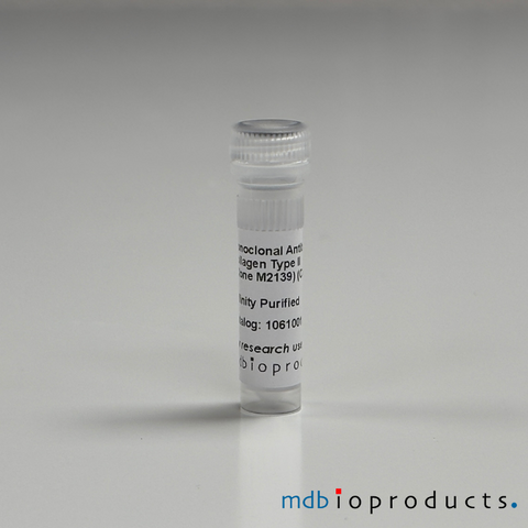
Collagen Type II, Monoclonal Antibody (Clone M2139), 100ugThis antibody (clone 2139) recognizes the conformational J1 epitope on the triple-helical structure of the native Collagen Type II molecule. Th...
-
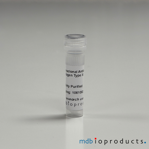
Collagen Type XI, Monoclonal Antibody, 100ugThis antibody (clone L10D9) binds to the triple-helical D3 epitope on collagen XI, which is shared with Collagen type II. The antibody binds to...
-
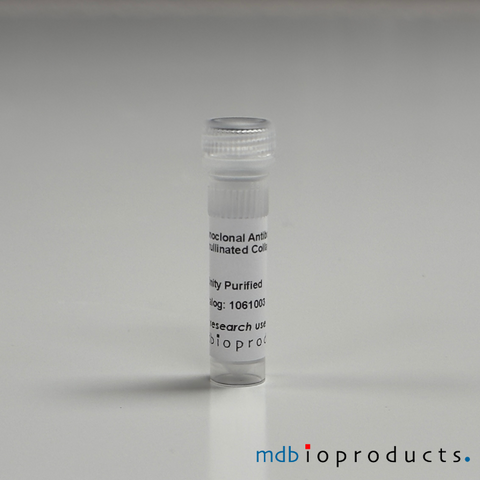
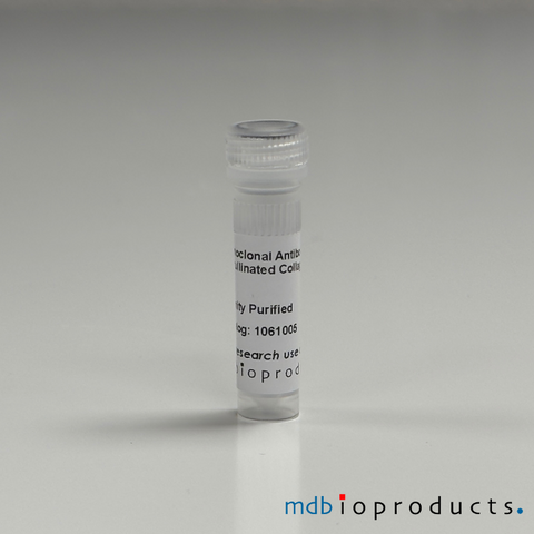
ACC1, Monoclonal Antibody, 100ugThis antibody binds ACC1 to the citrullinated triple-helical collagen type II (CII) epitope (position 359-369), the C1 epitope. The ACC1 antibo...
-
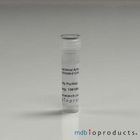
ACC4 (IgG1), Monoclonal Antibody, 100ugThis antibody is an anti-citrullinated protein antibody (ACPA) that, on Collagen alpha-1 (II) chain, binds specifically to citrullinated C1 epi...
-
ACC4 (IgG2b), Monoclonal Antibody, 100ugThis antibody is an anti-citrullinated protein antibody (ACPA) that on Collagen alpha-1 (II) chain binds specifically to citrullinated C1 epito...
-
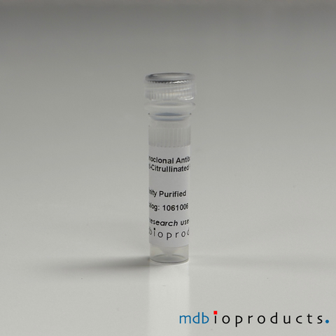
ACPA, Monoclonal Antibody (Clone E4NG), 100ugThis antibody recognizes multiple citrullinated proteins/peptides, including CCP2, citrullinated collagen type 2 (COL2) peptides, citrullinated...
-
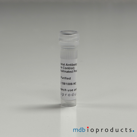
ACPA (Negative Control), Monoclonal Antibody (Clone E4NG-Mutant), 100ugThis antibody is a negative control for the E4NG antibody (product number 1061006). It is the mutated version of the E4NG antibody, with identi...
-
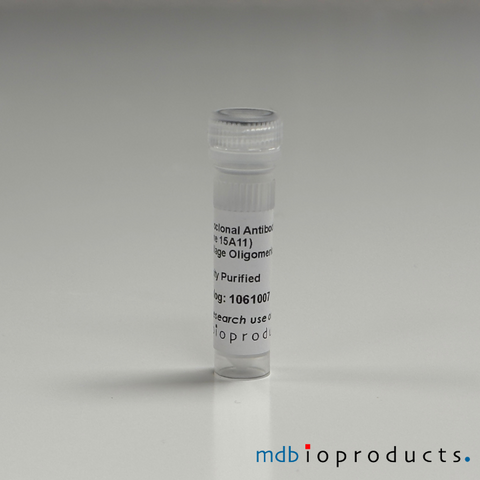
Cartilage Oligomeric Matrix Protein (COMP) , Monoclonal Antibody (Clone 15A11), 100ugThis antibody binds to the fourth EGF (Epithelial Growth Factor) -like domain of mouse COMP, comprising residues 232–252, called the P6 epitope...
-
Cartilage Oligomeric Matrix Protein (COMP), Monoclonal Antibody (Clone 16B5), 100ugThis antibody binds to the coiled-coil domain or pentamer COMP. It cross-reacts with mouse, rat and human. Cartilage Oligomeric Matrix Protein ...
-
Collagen Type II, Monoclonal Antibody (Clone CIIC1), 100ugThis antibody binds specifically to the C1 epitope on triple helica collagen type 2. It cross-reacts with mouse, rat, chicken, baboon and horse...
-
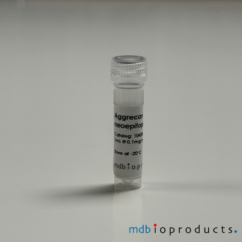
Aggrecan Antibody, C-terminal neoepitope NITEGE, 100 ugAggrecan monoclonal antibody to C-terminal neoepitope NITEGE (mouse, clone BC-13). This aggrecan degradation product usually remains within the tis...
-
Aggrecan Antibody, N-terminal neoepitope ARG, 100 ugAggrecan monoclonal antibody to N-terminal neoepitope ARG (mouse, clone BC-3). Aggrecan degradation products containing this neoepitope are rapidl...
-
Aggrecan Antibody, N-terminal neoepitope DIPEN, 100ugAggrecan monoclonal antibody to N-terminal neoepitope DIPEN (mouse, clone BC-4). Proteoglycans are categorized depending upon the nature of their g...
-
Aggrecan Antibody, N-terminal neoepitope FFGV, 100 ugAggrecan monoclonal antibody to N-terminal neoepitope FFGV (mouse, clone BC14). This fragment is rapidly released from the tissue when MMP cataboli...
-
Aggrecan IGD Antibody, 100 ugAggrecan IGD monoclonal antibody (mouse, clone 6B4). This antibody detects aggrecan metabolites (intact or matrix protease-catabolised) in human sy...
-
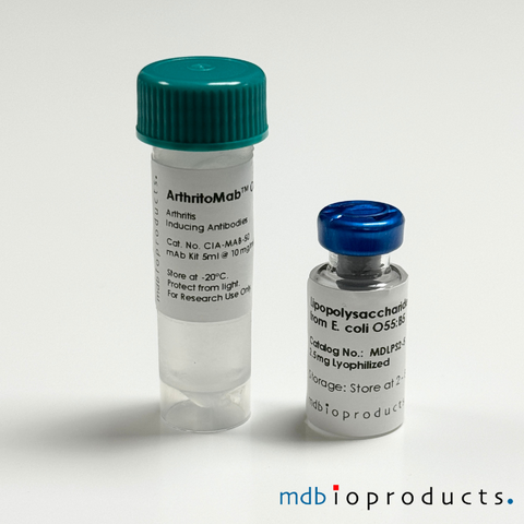
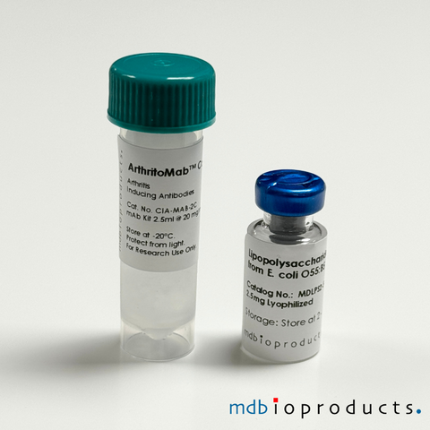
ArthritoMab™ Antibody Cocktail for Balb/c, DBA/1, R10.RIII, 50 mgArthritoMab™ Arthritis Inducing Antibody Cocktail is a cocktail of 4 arthritogenic monoclonal antibodies to collagen II (CII) used for inducing art...
-
ArthritoMab™ Antibody Cocktail for C57BL/6, TG, 50 mgArthritoMab™ Antibody Cocktail is a reformulated cocktail of 4 monoclonal antibodies for the induction of arthritis as an alternative to the widely...
-
Cartilage Link Protein, Antibody, 100 ugCartilage-link protein (HAPLN1) monoclonal antibody (clone 8A4). Cartilage-link protein (LP) is a glycoprotein present in cartilage that stabilizes...
-
Chondroitin Sulphate Neoepitope Antibody, 100ugChondroitin sulfate monoclonal antibody (mouse, clone 1B5) to detect the zero sulphated Chondroitin Sulphate stub neoepitope generated by chondroit...
-
Chondroitinase generated C-4-S & DS Antibody, 100ugChondroitin-4-sulfate (C-4-S) monoclonal antibody that's also specific to dermatan-sulfate (DS) neoepitope (mouse, clone 2B6). Monoclonal antibod...
-
Collagen Type I Antibody, anti-Mouse, 100 uLCollagen type I polyclonal antibody (rabbit anti-mouse) purified from rabbits injected with type I collagen that was extracted/purified from mouse ...
-
Collagen Type I Antibody, anti-Rat, 100 uLCollagen type I polyclonal antibody (rabbit anti-rat) purified from rabbits injected with type I collagen that was extracted/purified from rat skin...
-
Collagen Type II Antibody, anti-Human, 100 uLCollagen Type II polyclonal antibody (rabbit anti-human) purified from rabbits injected with type II collagen that was extracted/purified from huma...
-
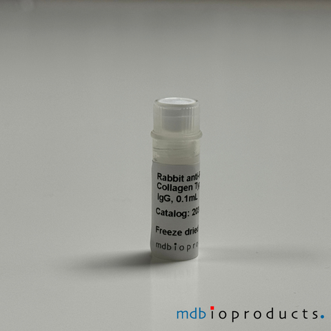
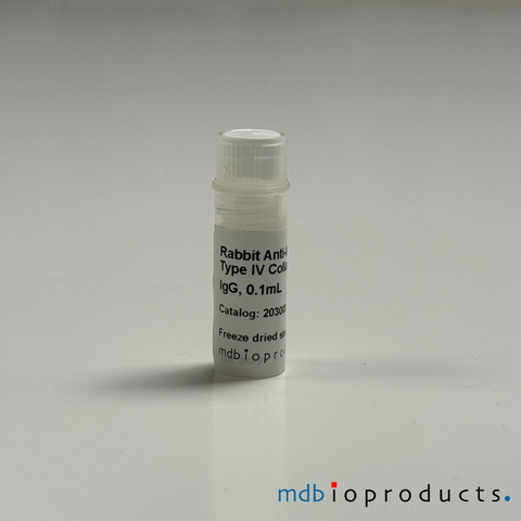
Collagen Type IV Antibody, anti-Mouse, 100 uLCollagen Type IV polyclonal antibody (rabbit anti-mouse) purified from rabbits injected with type IV collagen that was extracted/purified from mous...
-
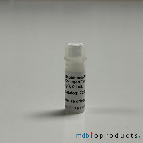
Collagen Type IV Antibody, anti-Rat, 100 uLAffinity chromatography purified rabbit antibody to rat type IV collagen extracted, purified from rodent tumor tissues. Purified, freeze-dried (0.5...
-
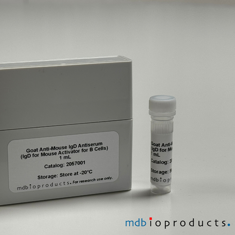
Goat anti-Mouse IgD, Antiserum, 1 mLImmunoglobulin D activator for B-cells. Preservative-free for in vivo application. Immunoglobulin D (IgD) is an antibody isotype that is found prim...
-
Hyaluronic Acid Binding Region, Antibody, 100 ugHyaluronic acid binding region (HABR), clone 1C6, monoclonal antibody is used to detect the HA-binding region of Aggrecan. Clear liquidy, 0.1 mg/mL...
-
Keratan Sulfate Antibody, 100 ugKeratan sulfate (KS) monoclonal antibody (mouse, clone 5D4) used to detect KS type I epitopes and KS type II epitopes. Liquid. Store at -20° C. K...
-
Lubricin Antibody, Bovine, 100 ugLubricin (PRG4) monoclonal antibody (mouse, clone 6A1) to detect superficial zone protein (SZP) from bovine articular cartilage and a non conformat...
-
Lubricin Antibody, Native Bovine, 100 ugLubricin (PRG4) monoclonal antibody (mouse, clone 3A4) to detect the native form of bovine lubricin. Does not recognize reduced or denatured lubric...
-
Lumican, Antibody, 100 ugLumican (LUM) monoclonal antibody (mouse, clone Lum-1) to detect a protein epitope in lumican. Liquid, 1 mL/vial. Concentration: 0.1 mg/mL Lumica...
-
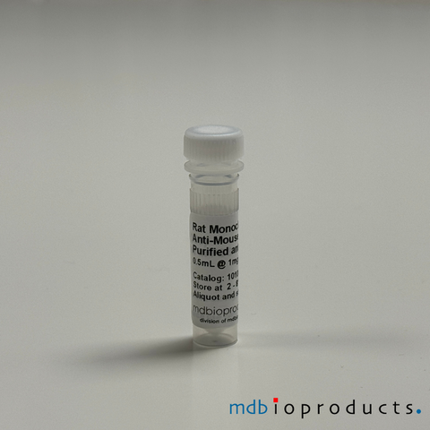
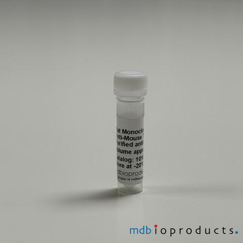
T1/ST2 (IL-33 R) Monoclonal Antibody, azide-free, 0.5 mLMouse T1/ST2 (IL-33 R) monoclonal antibody (Clone DJ8, Host / Isotype Subclass: Rat IgG1, light chain not isotyped), azide-free, for the identific...
-
T1/ST2 (IL-33 R) Mouse, Monoclonal Antibody, 0.5 mLMouse T1/ST2 (IL-33 R) monoclonal antibody (Clone: DJ8, Host / Isotype Subclass: Rat IgG1, light chain not isotyped) for the identification and pur...
-
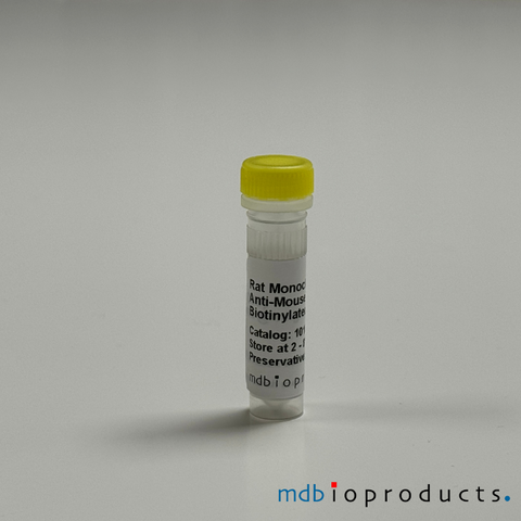
T1/ST2 (IL-33 R) Mouse, Monoclonal Antibody, Biotinylated, 0.5 mLMouse T1/ST2 (IL-33 R) biotinylated monoclonal antibody (Clone: DJ8, Host / Isotype Subclass: Rat IgG1, light chain not isotyped) for the identific...
-
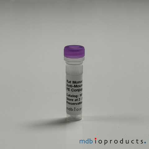
T1/ST2 (IL-33 R) Mouse, Monoclonal Antibody, PE Conjugated, 0.1 mLMouse T1/ST2 (IL-33 R) PE conjugated monoclonal antibody (Clone DJ8, Host / Isotype Subclass: Rat IgG1, light chain not isotyped) for the identific...
-
T1/ST2 (IL-33R) Mouse, Monoclonal Antibody, FITC, 0.5 mLMouse T1/ST2 (IL-33 R) FITC conjugated monoclonal antibody (Clone: DJ8, Host / Isotype Subclass: Rat IgG1, light chain not isotyped) for the identi...