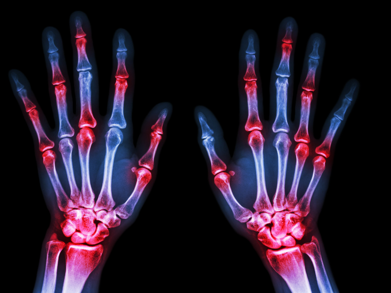
Collagen Induced Arthritis: an Experimental Model for Rheumatoid Arthritis (RA)
Sep 10 , 2020
Rodent models of immune-mediated arthritis (RMIA) are commonly used to evaluate the mechanisms of inflammatory joint disease, as well as test the efficacy of anti-arthritic compounds. The novel and rapid collagen antibody-induced arthritis (CAIA) model using the *ArthritoMab™ Antibody Cocktail for inducing arthritis within a short time (24-48 hrs), the classical models; Collagen induced arthritis (CIA), and the adjuvant induced arthritis (AIA) are the three commonly used models for this purpose.
Collagen Induced Arthritis (CIA) model
The Collagen Induced Arthritis (CIA) model is a commonly used 42 day model as it shares immunological and pathological similarities to human RA. Arthritis is initiated by intradermal injections of Collagen Type II (CII) emulsified in Complete Freund's Adjuvant (CFA). This causes an immune response generating antibodies to Collagen Type II. Therefore there is both a T cell and B cell component to the pathology. The joint destruction in collagen-induced arthritis involves cartilage destruction, bone resporption, synovial hyperplasia and periarticular cell infiltration thereby sharing many similarities to human RA.
Image: Longitudinal Section of the Paw from CIA Mice Model (H&E Staining). Source: MDB Laboratories

A-F (Vehicle Group): Severe lesions are presented in Figure A. in the tarsus (T) and in the digits (D). The boxed areas are shown in Figure B. (digits) and Figure C. (tarsus). Boxed areas in C. are presented at higher magnification in Figures D-F. There is severe inflammatory infiltration, fibrosis with loss of cartilage, and widespread destruction of tarsal and digits bones (arrows) as well as soft tissue. Many of the infiltrating cells are neutrophils.G-K (Dexamethasone Group) The tarsus is within normal limits and is shown at higher magnification in Figures H, I and J. The bones are intact (H,I) and joint spaces are clearly identified as well as optically empty (J). Cartilage is spanned by double arrowheads (K).
Image: Sections of Paw from CIA Model Stained for Macrophage IHC Marker CD206. Source MDB Labs

Figure A. Medium magnification of paw section including CD206+ cells in infiltrates within soft tissue (arrows) and in bone marrow (circle). Figure B. High magnification of CD206+ cells in bone marrow.
Image: Paw Thickness & Clinical Scores. Source MDB Labs.

Representative data from the mouse Collagen-Induced Arthritis (CIA) study. DBA/1 mice were induced with type II Collagen and given a boost on day 21. Data shows average paw thickness in mm (top) and total arthritis clinical scores (bottom) for naive, vehicle, and Dexamethasone treatment groups.
Products for Collagen-Induced Arthritis Model (CIA):
MD Biosciences offers highly purified (>99%), quality immunization grade Collagen Type II products are very popular among researchers worldwide. Academic, pharmaceutical, biotech, and governmental laboratories utilize our Bovine or Chicken Collagen Type II for their arthritis work (Balb/c, DBA, C57BL/6).
Our Collagen Type II is a preferred reagent for the Collagen Induced Arthritis (CIA) Model due to its high quality and purified material (99%, as determined by SDS-PAGE) and it enables researchers to induce a high incidence and severity in the disease model.