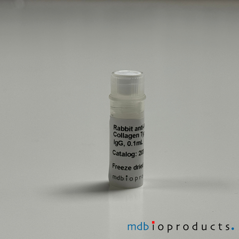
Collagen Type I Antibody, anti-Mouse, 100 uL
Collagen type I polyclonal antibody (rabbit anti-mouse) purified from rabbits injected with type I collagen that was extracted/purified from mouse skin. Purified, freeze-dried antibody in a 0.1 mL vial. Type I...
Antibodies
203002-1
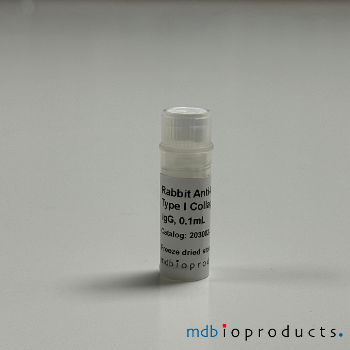
Collagen type I polyclonal antibody (rabbit anti-mouse) purified from rabbits injected with type I collagen that was extracted/purified from mouse skin. Purified, freeze-dried antibody in a 0.1 mL vial.
Type I collagen is the most abundant form of collagen in the human body and is synthesized mainly by fibroblasts, osteoblasts, odontoblasts and chondroblasts. It is located in the extracellular matrix of many tissues of the body including cartilage, bone, tendon, skin and the sclera of the eye. Type I collagen is composed of two pro-_1(I) chains, produced from the COL1A1 gene, and one pro-_2(I) chain, produced from the COL1A2 gene. Mutations in the genes that produce collagen type I are responsible for causing various health conditions including Ehlers-Danlos syndrome, osteogenesis imperfecta, osteoporosis and Caffey disease.
References/Citations:
Borza, C. M., Bolas, G., Bock, F., Zhang, X., Akabogu, F. C., Zhang, M. Z., ... & Pozzi, A. (2022). DDR1 contributes to kidney inflammation and fibrosis by promoting the phosphorylation of BCR and STAT3. JCI insight, 7(3).
Lu, C. L., Cain, J., Brudvig, J., Ortmeier, S., Boyadjiev, S. A., Weimer, J. M., & Kim, J. (2021). Collagen has a unique SEC24 preference for efficient export from the endoplasmic reticulum. bioRxiv.
Bota-Rabassedas, N., Guo, H. F., Banerjee, P., Chen, Y., Terajima, M., Yamauchi, M., & Kurie, J. M. (2020). Use of osteoblast-derived matrix to assess the influence of collagen modifications on cancer cells. Matrix Biology Plus, 8, 100047.
Saraswati, S., Lietman, C. D., Li, B., Mathew, S., Zent, R., & Young, P. P. (2020). Small proline-rich repeat 3 is a novel coordinator of PDGFRβ and integrin β1 crosstalk to augment proliferation and matrix synthesis by cardiac fibroblasts. The FASEB Journal.
Feng, Y., Li, M., Wang, S., Cong, W., Hu, G., Song, Y., ... & Zhang, Y. (2020). Paired box 6 inhibits cardiac fibroblast differentiation. Biochemical and Biophysical Research Communications, 528(3), 561-566.
Saraswati, S., Marrow, S. M., Watch, L. A., & Young, P. P. (2019). Identification of a pro-angiogenic functional role for FSP1-positive fibroblast subtype in wound healing. Nature Communications, 10(1), 1-16.
Nawaito, S. A., Sahadevan, P., Sahmi, F., Gaestel, M., Calderone, A., & Allen, B. G. (2019). Transcript levels for extracellular matrix proteins are altered in MK5-deficient cardiac ventricular fibroblasts. Journal of Molecular and Cellular Cardiology, 132, 164-177.
Pesevski, Z., Kvasilova, A., Stopkova, T., Nanka, O., Drobna Krejci, E., Buffinton, C., ... & Sedmera, D. (2018). Endocardial fibroelastosis is secondary to hemodynamic alterations in the chick embryonic model of hypoplastic left heart syndrome. Developmental Dynamics, 247(3), 509-520.
Surinkaew, S., Aflaki, M., Takawale, A., Chen, Y., Qi, X. Y., Gillis, M. A., ... & Nattel, S. (2018). Exchange protein activated by cyclic-adenosine monophosphate (Epac) regulates atrial fibroblast function and controls cardiac remodelling. Cardiovascular research, 115(1), 94-106.
Viquez, O. M., Yazlovitskaya, E. M., Tu, T., Mernaugh, G., Secades, P., McKee, K. K., ... & Gewin, L. C. (2017). Integrin alpha6 maintains the structural integrity of the kidney collecting system. Matrix Biology, 57, 244-257.
Feng, Y., Wang, S., Zhang, Y., & Xiao, H. (2017). Metformin attenuates renal fibrosis in both AMPK α2‐dependent and independent manners. Clinical and Experimental Pharmacology and Physiology, 44(6), 648-655.
Chen, X., & Thibeault, S. L. (2016). Cell–cell interaction between vocal fold fibroblasts and bone marrow mesenchymal stromal cells in three‐dimensional hyaluronan hydrogel. Journal of tissue engineering and regenerative medicine, 10(5), 437-446.
Dupuis, L. E., Doucette, L., Rice, A. K., Lancaster, A. E., Berger, M. G., Chakravarti, S., & Kern, C. B. (2016). Development of myotendinous‐like junctions that anchor cardiac valves requires fibromodulin and lumican. Developmental Dynamics, 245(10), 1029-1042.
Seet, L. F., Toh, L. Z., Finger, S. N., Chu, S. W., Stefanovic, B., & Wong, T. T. (2016). Valproic acid suppresses collagen by selective regulation of Smads in conjunctival fibrosis. Journal of Molecular Medicine, 94(3), 321-334.
Pankova, D., Chen, Y., Terajima, M., Schliekelman, M. J., Baird, B. N., Fahrenholtz, M., ... & Ahn, Y. H. (2016). Cancer-associated fibroblasts induce a collagen cross-link switch in tumor stroma. Molecular Cancer Research, 14(3), 287-295.
Neelisetty, S., Alford, C., Reynolds, K., Woodbury, L., Nlandu-khodo, S., Yang, H., ... & Gewin, L. (2015). Renal fibrosis is not reduced by blocking transforming growth factor-β signaling in matrix-producing interstitial cells. Kidney international, 88(3), 503-514.
Wang, H., Chen, X., Su, Y., Paueksakon, P., Hu, W., Zhang, M. Z., ... & Pozzi, A. (2015). p47phox contributes to albuminuria and kidney fibrosis in mice. Kidney international, 87(5), 948-962.
Singh, S. P., Tao, S., Fields, T. A., Webb, S., Harris, R. C., & Rao, R. (2015). Glycogen synthase kinase-3 inhibition attenuates fibroblast activation and development of fibrosis following renal ischemia-reperfusion in mice. Disease models & mechanisms, 8(8), 931-940.
Trombetta‐eSilva, J., Rosset, E. A., Hepfer, R. G., Wright, G. J., Baicu, C., Yao, H., & Bradshaw, A. D. (2015). Decreased Mechanical Strength and Collagen Content in SPARC‐Null Periodontal Ligament Is Reversed by Inhibition of Transglutaminase Activity. Journal of bone and mineral research, 30(10), 1914-1924.
Zhu, M., Tao, J., Vasievich, M. P., Wei, W., Zhu, G., Khoriaty, R. N., & Zhang, B. (2015). Neural tube opening and abnormal extraembryonic membrane development in SEC23A deficient mice. Scientific reports, 5, 15471.
Manley Jr, E., Perosky, J. E., Khoury, B. M., Reddy, A. B., Kozloff, K. M., & Alford, A. I. (2015). Thrombospondin-2 deficiency in growing mice alters bone collagen ultrastructure and leads to a brittle bone phenotype. Journal of Applied Physiology, 119(8), 872-881.
Sochman, J., Peregrin, J. H., Pavcnik, D., Uchida, B. T., Timmermans, H. A., Sedmera, D., ... & Rosch, J. (2014). Reverse endoventricular artificial obturator in tricuspid valve position. Experimental feasibility research study. Physiological research, 63(2), 157.
Bohuslavova, R., Kolar, F., Sedmera, D., Skvorova, L., Papousek, F., Neckar, J., & Pavlinkova, G. (2014). Partial deficiency of HIF-1α stimulates pathological cardiac changes in streptozotocin-induced diabetic mice. BMC endocrine disorders, 14(1), 11.
Chen, X., Wang, H., Liao, H. J., Hu, W., Gewin, L., Mernaugh, G., ... & Fässler, R. (2014). Integrin-mediated type II TGF-β receptor tyrosine dephosphorylation controls SMAD-dependent profibrotic signaling. The Journal of clinical investigation, 124(8), 3295-3310.
Zimmerman, K. A., Graham, L. V., Pallero, M. A., & Murphy-Ullrich, J. E. (2013). Calreticulin (CRT) regulates Transforming Growth Factor-β (TGF-β) stimulated extracellular matrix production. Journal of Biological Chemistry, jbc-M112.
Rosa, R. G., Akgul, Y., Joazeiro, P. P., & Mahendroo, M. (2012). Changes of large molecular weight hyaluronan and versican in the mouse pubic symphysis through pregnancy. Biology of reproduction, 86(2).
Baicu, C. F., Zhang, Y., Van Laer, A. O., Renaud, L., Zile, M. R., & Bradshaw, A. D. (2012). Effects of the absence of procollagen C-endopeptidase enhancer-2 on myocardial collagen accumulation in chronic pressure overload. American Journal of Physiology-Heart and Circulatory Physiology, 303(2), H234.
Dawson, K., Wu, C. T., Qi, X. Y., & Nattel, S. (2012). Congestive heart failure effects on atrial fibroblast phenotype: differences between freshly-isolated and cultured cells. PLoS One, 7(12), e52032.
Chen, J., Chen, J. K., Nagai, K., Plieth, D., Tan, M., Lee, T. C., ... & Harris, R. C. (2012). EGFR signaling promotes TGFβ-dependent renal fibrosis. Journal of the American Society of Nephrology, 23(2), 215-224.
Dagher, P. C., Mai, E. M., Hato, T., Lee, S. Y., Anderson, M. D., Karozos, S. C., ... & Sutton, T. A. (2011). The p53 inhibitor pifithrin-α can stimulate fibrosis in a rat model of ischemic acute kidney injury. American Journal of Physiology-Renal Physiology, 302(2), F284-F291.
Akins, M. L., Luby-Phelps, K., Bank, R. A., & Mahendroo, M. (2011). Cervical softening during pregnancy: regulated changes in collagen cross-linking and composition of matricellular proteins in the mouse. Biology of reproduction, 84(5), 1053-1062.
Harris, B. S., Zhang, Y., Card, L., Rivera, L. B., Brekken, R. A., & Bradshaw, A. D. (2011). SPARC regulates collagen interaction with cardiac fibroblast cell surfaces. American Journal of Physiology-Heart and Circulatory Physiology, 301(3), H841-H847.
Graham, L. V. D., Sweetwyne, M. T., Pallero, M. A., & Murphy-Ullrich, J. E. (2010). Intracellular calreticulin regulates multiple steps in fibrillar collagen expression, trafficking, and processing into the extracellular matrix. Journal of Biological Chemistry, 285(10), 7067-7078.
He, W., Wang, Y., Zhang, M. Z., You, L., Davis, L. S., Fan, H., ... & Hao, C. M. (2010). Sirt1 activation protects the mouse renal medulla from oxidative injury. The Journal of clinical investigation, 120(4), 1056-1068.
Cervical Softening During Pregnancy: Regulated Changes in Collagen Cross-Linking and Composition of Matricellular Proteins in the Mouse.Meredith L. Akins, Katherine Luby-Phelps, Ruud A. Bank, and Mala Mahendroo Biol Reprod, May 2011; 84: 1053 - 1062Intracellular Calreticulin Regulates Multiple Steps in Fibrillar Collagen Expression, Trafficking, and Processing into the Extracellular Matrix
Lauren Van Duyn Graham, et al. J. Biol. Chem., Mar 2010; 285: 7067 - 7078.Sirt1 activation protects the mouse renal medulla from oxidative injury.
He W, et al. J Clin Invest. 2010 Apr; 120(4):1056-68.Type XIV collagen regulates fibrillogenesis: premature collagen fibril growth and tissue dysfunction in null mice.
Ansorge HL, et al. J Biol Chem. Mar 2009; 284(13): 8427-38.SPARC Regulates Processing of Procollagen I and Collagen Fibrillogenesis in Dermal Fibroblasts
Tyler J. Rentz et al., J. Biol. Chem., Jul 2007; 282: 22062 - 22071.
The Calreticulin-Binding Sequence of Thrombospondin 1 Regulates Collagen Expression and Organization During Tissue Remodeling
Mariya T. Sweetwyne, Manuel A. Pallero, Ailing Lu, Lauren Van Duyn Graham, and Joanne E. Murphy-Ullrich
Am. J. Pathol., Oct 2010; 177: 1710 - 1724.
Product Insert (PDF) - Informational use only. Please refer to insert included with product.
Data/Specifications:
Format: Purified, freeze-dried antibody in 0.1 mL vial. Reconstitute with 0.1 mL distilled water and store aliquots at -20°C.
Specificity (% at 1:500 RIA dilution):
Mouse Collagen, Type I : 100%
Mouse Collagen, Types II, IV : < 0.1%
Mouse collagen type III: < 1.0%
Human, chicken, rat Collagen Type I: < 0.1%
Related Products
Additional products you might interested in.
-
Collagen Type II, Monoclonal Antibody (Clone M2139), 100ugThis antibody (clone 2139) recognizes the conformational J1 epitope on the triple-helical structure of the native Collagen Type II molecule. Th...
-
Collagen Type XI, Monoclonal Antibody, 100ugThis antibody (clone L10D9) binds to the triple-helical D3 epitope on collagen XI, which is shared with Collagen type II. The antibody binds to...
-
ACC1, Monoclonal Antibody, 100ugThis antibody binds ACC1 to the citrullinated triple-helical collagen type II (CII) epitope (position 359-369), the C1 epitope. The ACC1 antibo...
-
ACC4 (IgG1), Monoclonal Antibody, 100ugThis antibody is an anti-citrullinated protein antibody (ACPA) that, on Collagen alpha-1 (II) chain, binds specifically to citrullinated C1 epi...
-
ACC4 (IgG2b), Monoclonal Antibody, 100ugThis antibody is an anti-citrullinated protein antibody (ACPA) that on Collagen alpha-1 (II) chain binds specifically to citrullinated C1 epito...
-
ACPA, Monoclonal Antibody (Clone E4NG), 100ugThis antibody recognizes multiple citrullinated proteins/peptides, including CCP2, citrullinated collagen type 2 (COL2) peptides, citrullinated...
-
ACPA (Negative Control), Monoclonal Antibody (Clone E4NG-Mutant), 100ugThis antibody is a negative control for the E4NG antibody (product number 1061006). It is the mutated version of the E4NG antibody, with identi...
-
Cartilage Oligomeric Matrix Protein (COMP) , Monoclonal Antibody (Clone 15A11), 100ugThis antibody binds to the fourth EGF (Epithelial Growth Factor) -like domain of mouse COMP, comprising residues 232–252, called the P6 epitope...
-
Cartilage Oligomeric Matrix Protein (COMP), Monoclonal Antibody (Clone 16B5), 100ugThis antibody binds to the coiled-coil domain or pentamer COMP. It cross-reacts with mouse, rat and human. Cartilage Oligomeric Matrix Protein ...
-
Collagen Type II, Monoclonal Antibody (Clone CIIC1), 100ugThis antibody binds specifically to the C1 epitope on triple helica collagen type 2. It cross-reacts with mouse, rat, chicken, baboon and horse...
-
Aggrecan Antibody, C-terminal neoepitope NITEGE, 100 ugAggrecan monoclonal antibody to C-terminal neoepitope NITEGE (mouse, clone BC-13). This aggrecan degradation product usually remains within the tis...
-
Aggrecan Antibody, N-terminal neoepitope ARG, 100 ugAggrecan monoclonal antibody to N-terminal neoepitope ARG (mouse, clone BC-3). Aggrecan degradation products containing this neoepitope are rapidl...
-
Aggrecan Antibody, N-terminal neoepitope DIPEN, 100ugAggrecan monoclonal antibody to N-terminal neoepitope DIPEN (mouse, clone BC-4). Proteoglycans are categorized depending upon the nature of their g...
-
Aggrecan Antibody, N-terminal neoepitope FFGV, 100 ugAggrecan monoclonal antibody to N-terminal neoepitope FFGV (mouse, clone BC14). This fragment is rapidly released from the tissue when MMP cataboli...
-
Aggrecan IGD Antibody, 100 ugAggrecan IGD monoclonal antibody (mouse, clone 6B4). This antibody detects aggrecan metabolites (intact or matrix protease-catabolised) in human sy...
-
ArthritoMab™ Antibody Cocktail for Balb/c, DBA/1, R10.RIII, 50 mgArthritoMab™ Arthritis Inducing Antibody Cocktail is a cocktail of 4 arthritogenic monoclonal antibodies to collagen II (CII) used for inducing art...
-
ArthritoMab™ Antibody Cocktail for C57BL/6, TG, 50 mgArthritoMab™ Antibody Cocktail is a reformulated cocktail of 4 monoclonal antibodies for the induction of arthritis as an alternative to the widely...
-
Cartilage Link Protein, Antibody, 100 ugCartilage-link protein (HAPLN1) monoclonal antibody (clone 8A4). Cartilage-link protein (LP) is a glycoprotein present in cartilage that stabilizes...
-
Chondroitin Sulphate Neoepitope Antibody, 100ugChondroitin sulfate monoclonal antibody (mouse, clone 1B5) to detect the zero sulphated Chondroitin Sulphate stub neoepitope generated by chondroit...
-
Chondroitinase generated C-4-S & DS Antibody, 100ugChondroitin-4-sulfate (C-4-S) monoclonal antibody that's also specific to dermatan-sulfate (DS) neoepitope (mouse, clone 2B6). Monoclonal antibod...
-
Collagen Type I Antibody, anti-Rat, 100 uLCollagen type I polyclonal antibody (rabbit anti-rat) purified from rabbits injected with type I collagen that was extracted/purified from rat skin...
-
Collagen Type II Antibody, anti-Human, 100 uLCollagen Type II polyclonal antibody (rabbit anti-human) purified from rabbits injected with type II collagen that was extracted/purified from huma...
-
Collagen Type IV Antibody, anti-Mouse, 100 uLCollagen Type IV polyclonal antibody (rabbit anti-mouse) purified from rabbits injected with type IV collagen that was extracted/purified from mous...
-
Collagen Type IV Antibody, anti-Rat, 100 uLAffinity chromatography purified rabbit antibody to rat type IV collagen extracted, purified from rodent tumor tissues. Purified, freeze-dried (0.5...
-
Goat anti-Mouse IgD, Antiserum, 1 mLImmunoglobulin D activator for B-cells. Preservative-free for in vivo application. Immunoglobulin D (IgD) is an antibody isotype that is found prim...
-
Hyaluronic Acid Binding Region, Antibody, 100 ugHyaluronic acid binding region (HABR), clone 1C6, monoclonal antibody is used to detect the HA-binding region of Aggrecan. Clear liquidy, 0.1 mg/mL...
-
Keratan Sulfate Antibody, 100 ugKeratan sulfate (KS) monoclonal antibody (mouse, clone 5D4) used to detect KS type I epitopes and KS type II epitopes. Liquid. Store at -20° C. K...
-
Keratocan Antibody, 100 ugKeratocan (KERA) monoclonal antibody (mouse, clone Ker-1) to detect keratocan. 1 mL/vial. Concentration is 0.1 mg/mL. Normal keratocan expressio...
-
Lubricin Antibody, Bovine, 100 ugLubricin (PRG4) monoclonal antibody (mouse, clone 6A1) to detect superficial zone protein (SZP) from bovine articular cartilage and a non conformat...
-
Lubricin Antibody, Native Bovine, 100 ugLubricin (PRG4) monoclonal antibody (mouse, clone 3A4) to detect the native form of bovine lubricin. Does not recognize reduced or denatured lubric...
-
Lumican, Antibody, 100 ugLumican (LUM) monoclonal antibody (mouse, clone Lum-1) to detect a protein epitope in lumican. Liquid, 1 mL/vial. Concentration: 0.1 mg/mL Lumica...
-
T1/ST2 (IL-33 R) Monoclonal Antibody, azide-free, 0.5 mLMouse T1/ST2 (IL-33 R) monoclonal antibody (Clone DJ8, Host / Isotype Subclass: Rat IgG1, light chain not isotyped), azide-free, for the identific...
-
T1/ST2 (IL-33 R) Mouse, Monoclonal Antibody, 0.5 mLMouse T1/ST2 (IL-33 R) monoclonal antibody (Clone: DJ8, Host / Isotype Subclass: Rat IgG1, light chain not isotyped) for the identification and pur...
-
T1/ST2 (IL-33 R) Mouse, Monoclonal Antibody, Biotinylated, 0.5 mLMouse T1/ST2 (IL-33 R) biotinylated monoclonal antibody (Clone: DJ8, Host / Isotype Subclass: Rat IgG1, light chain not isotyped) for the identific...
-
T1/ST2 (IL-33 R) Mouse, Monoclonal Antibody, PE Conjugated, 0.1 mLMouse T1/ST2 (IL-33 R) PE conjugated monoclonal antibody (Clone DJ8, Host / Isotype Subclass: Rat IgG1, light chain not isotyped) for the identific...
-
T1/ST2 (IL-33R) Mouse, Monoclonal Antibody, FITC, 0.5 mLMouse T1/ST2 (IL-33 R) FITC conjugated monoclonal antibody (Clone: DJ8, Host / Isotype Subclass: Rat IgG1, light chain not isotyped) for the identi...